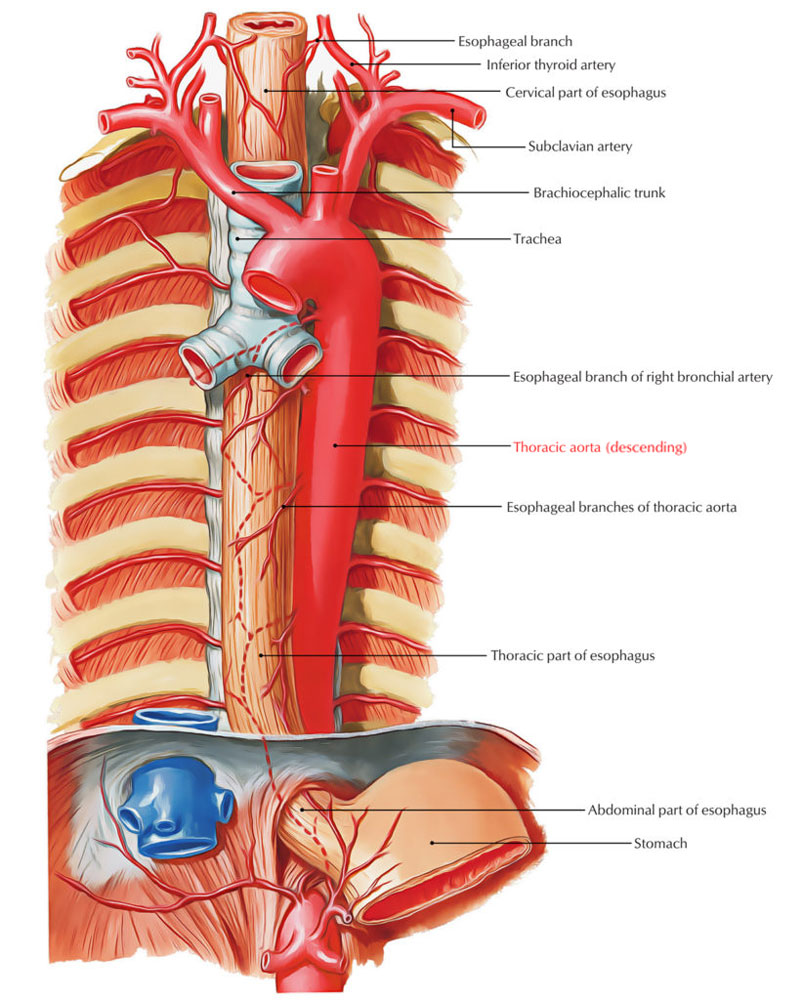
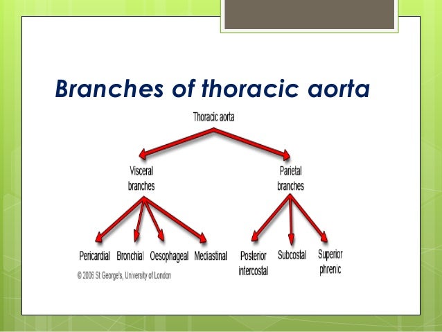
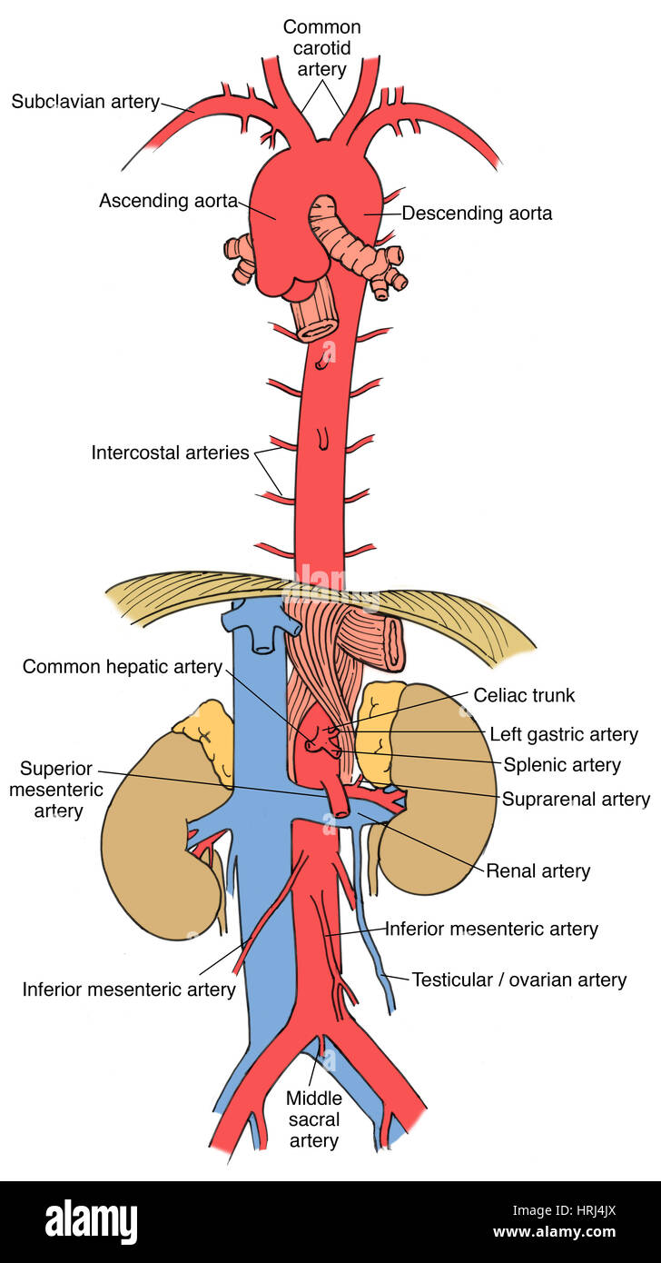
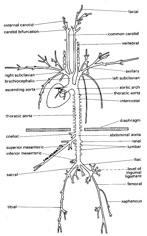
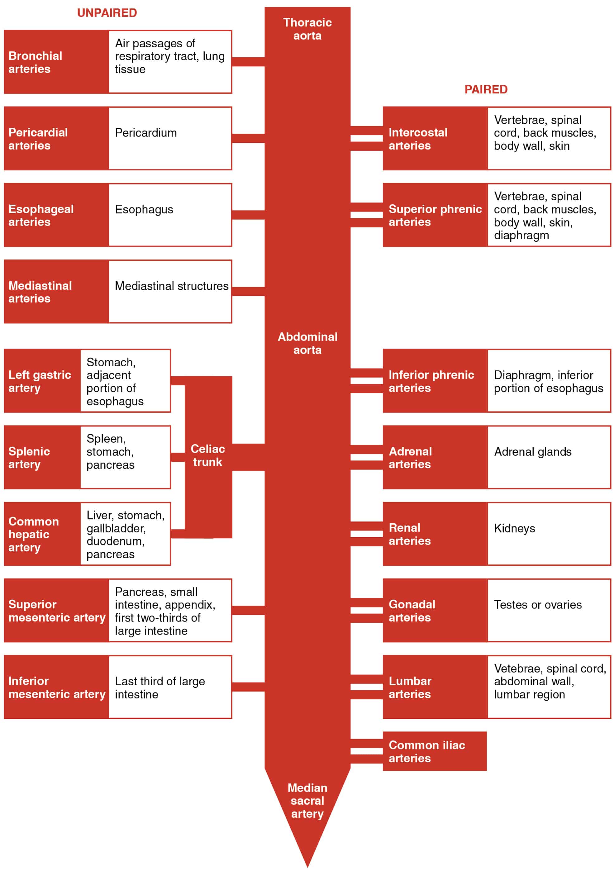
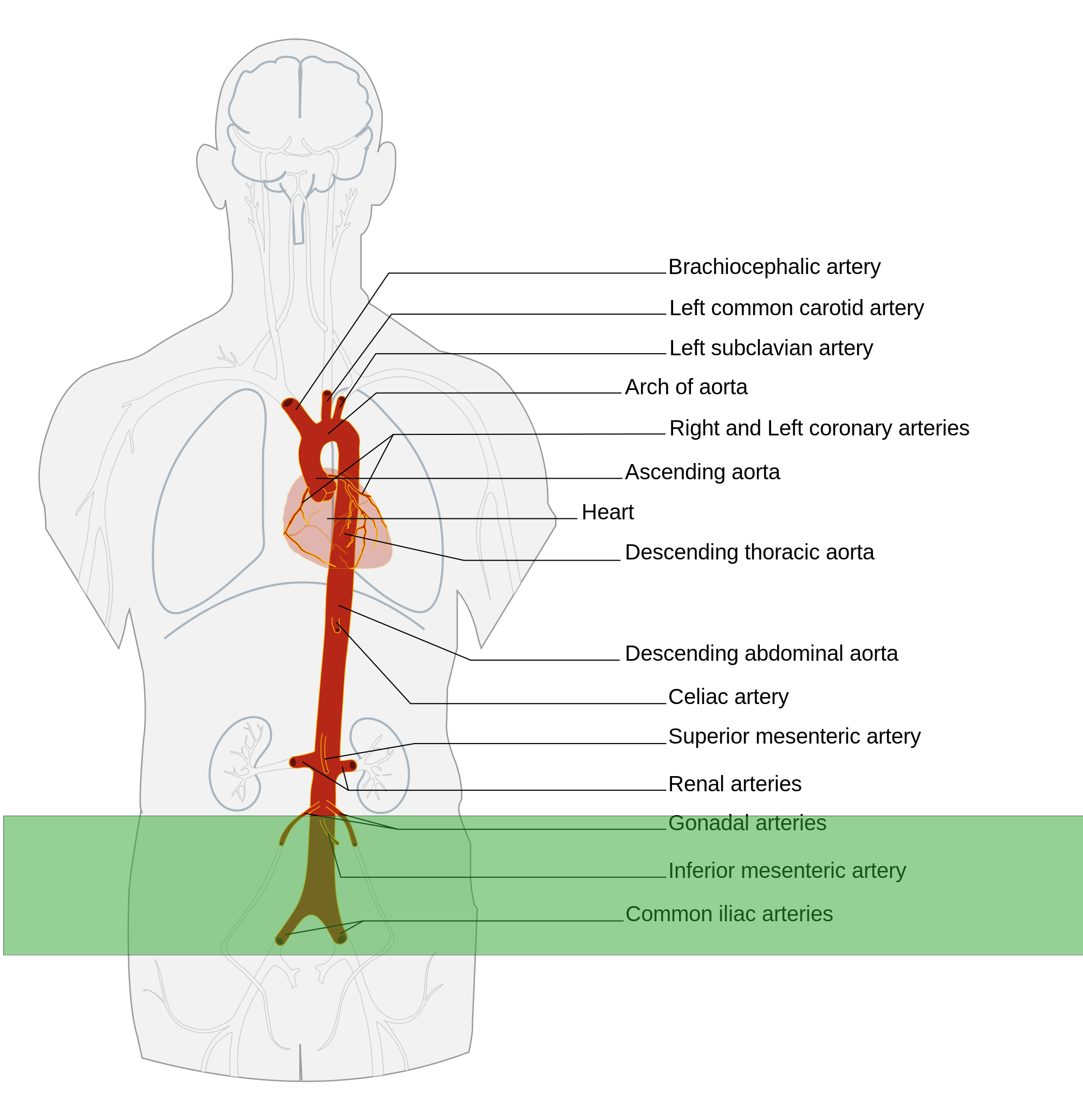
Thoracic Aorta Branches Diagram
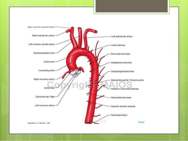
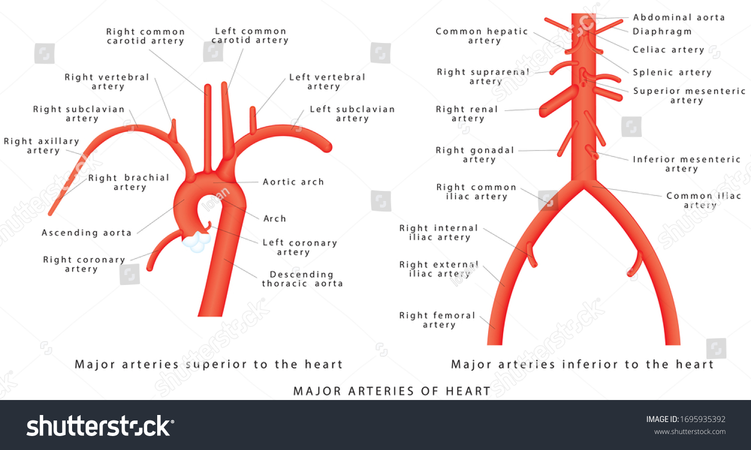
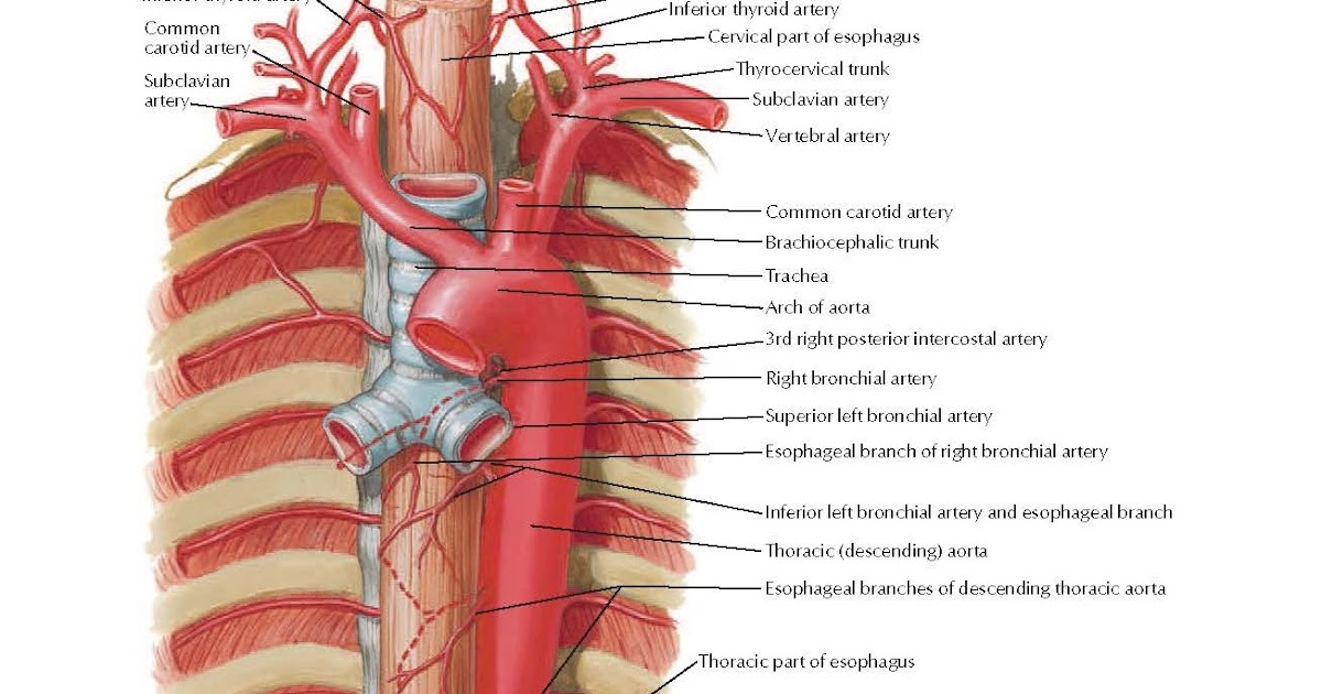
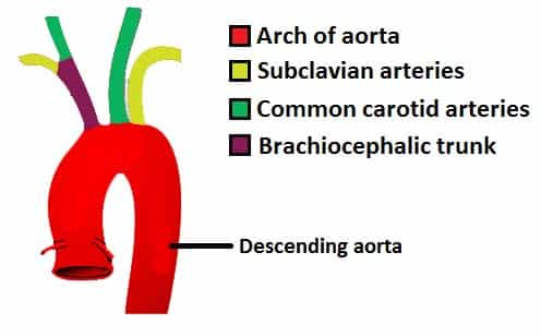
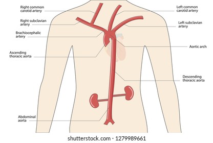
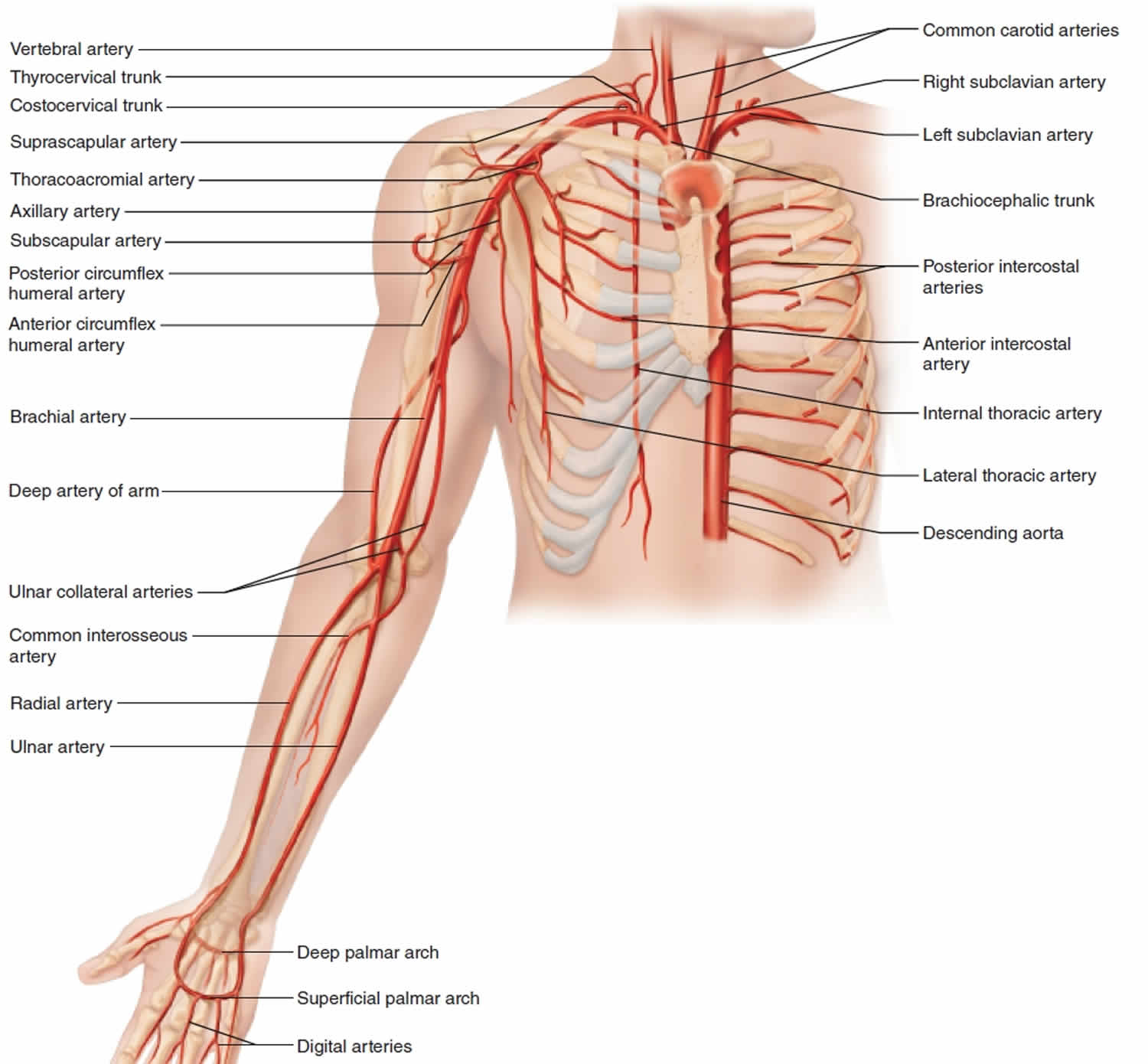
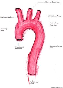
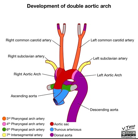
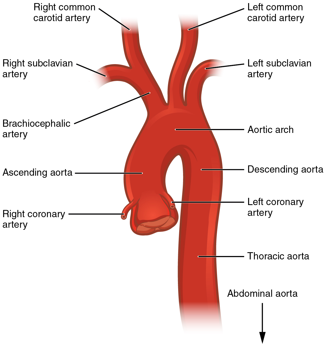
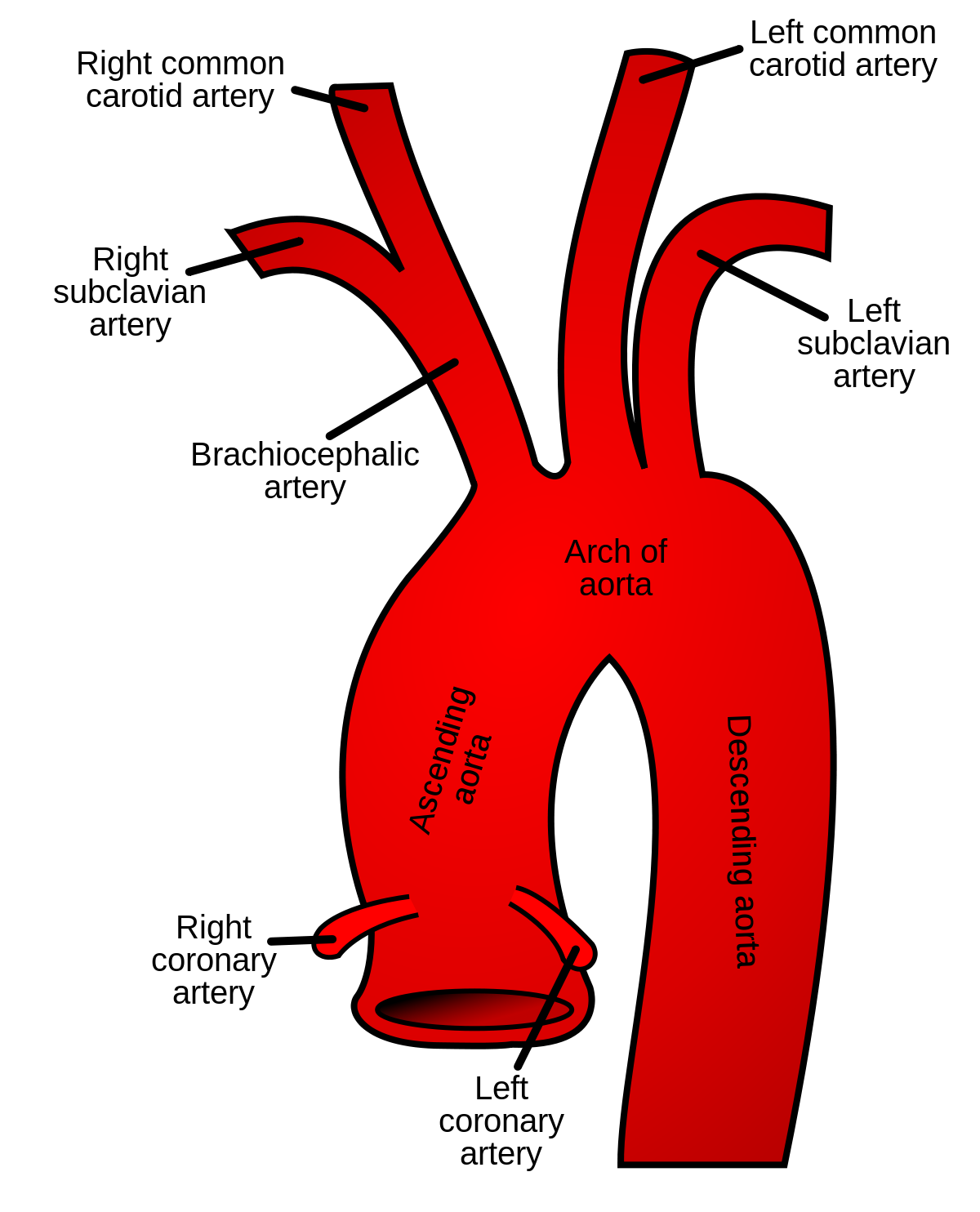
The first and largest branch is the brachiocephalic trunk.
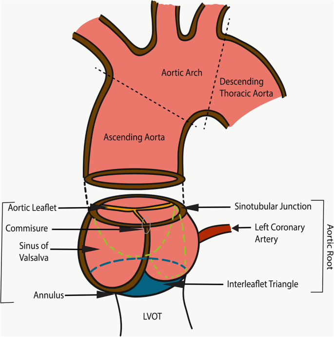
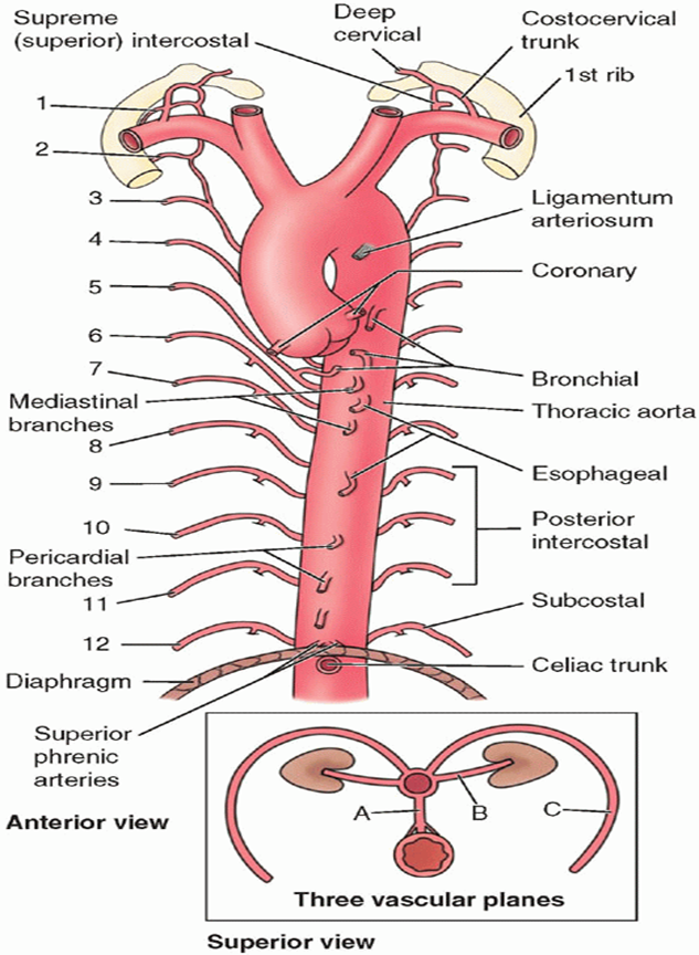
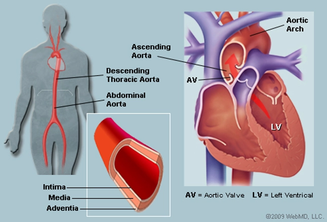
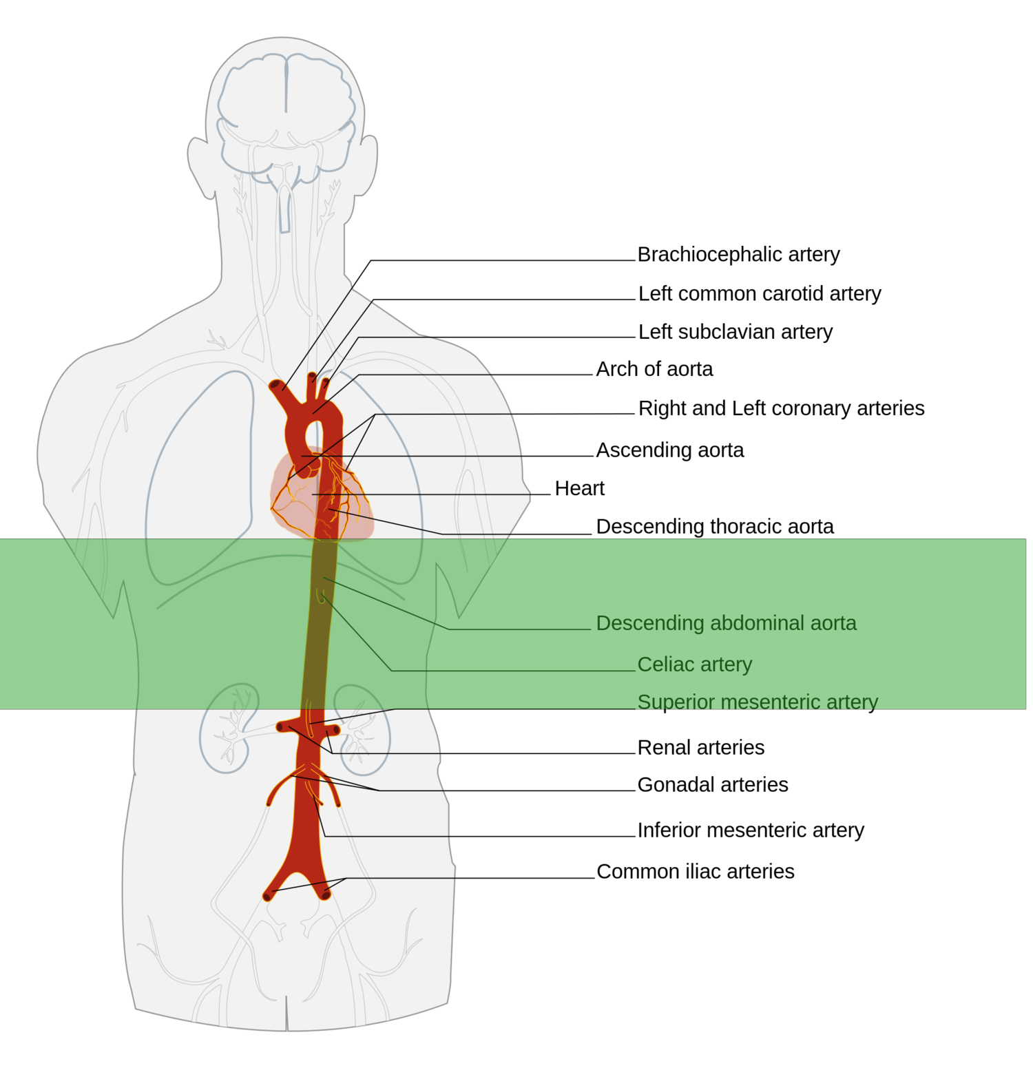
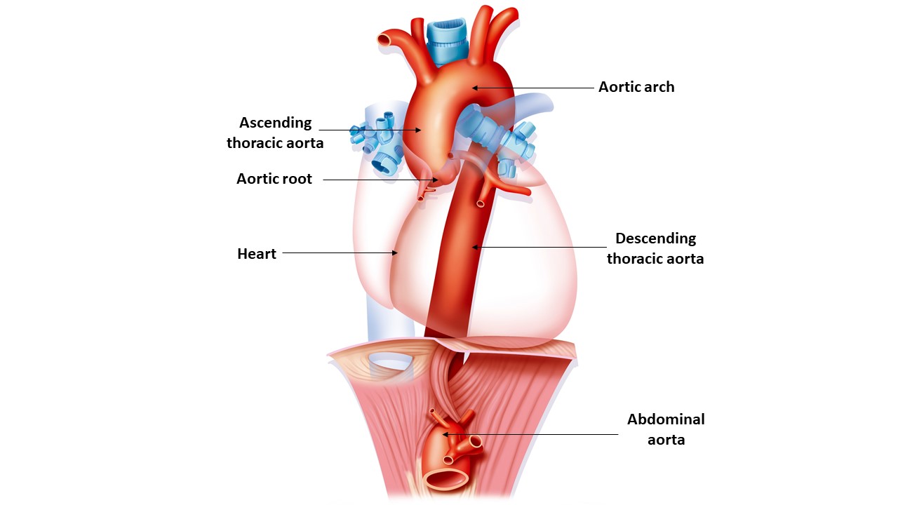
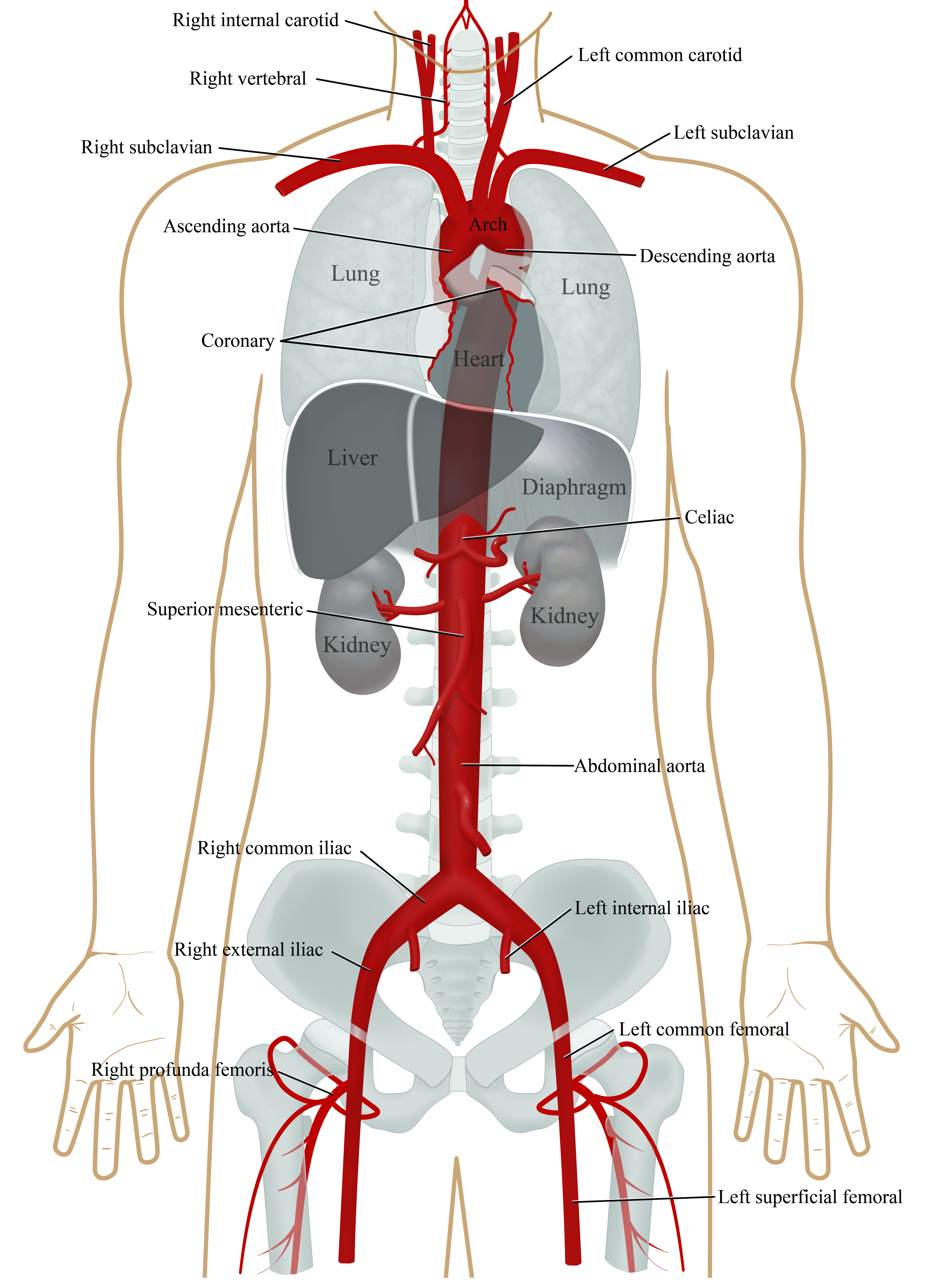
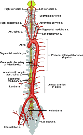
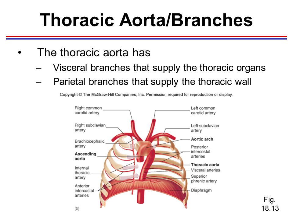
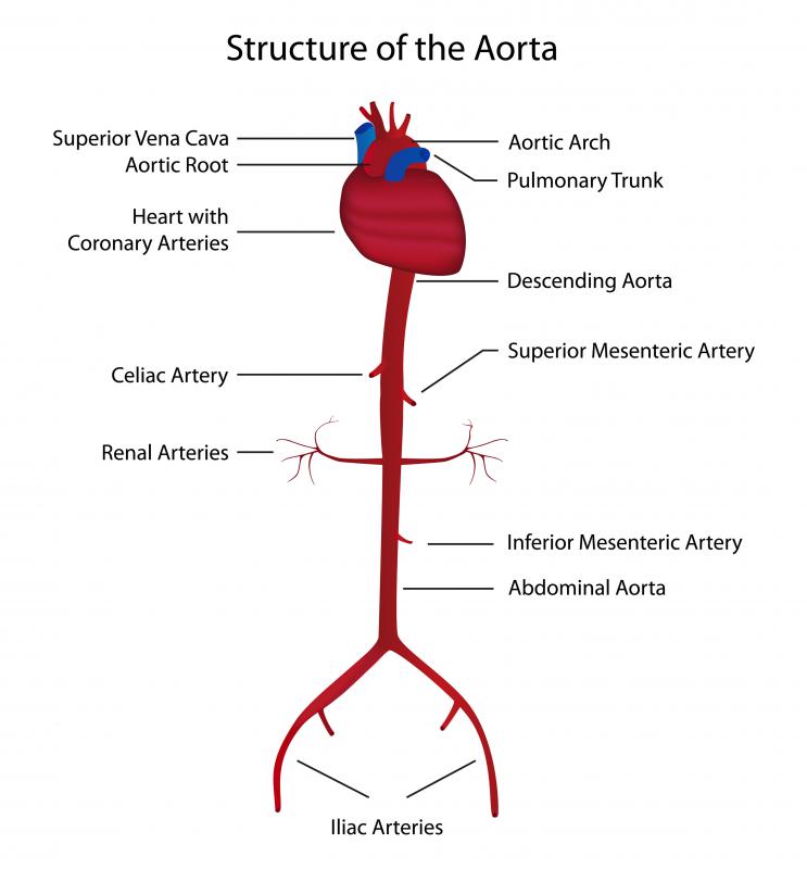
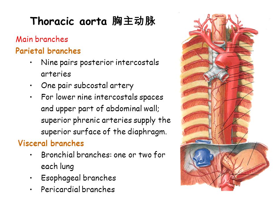
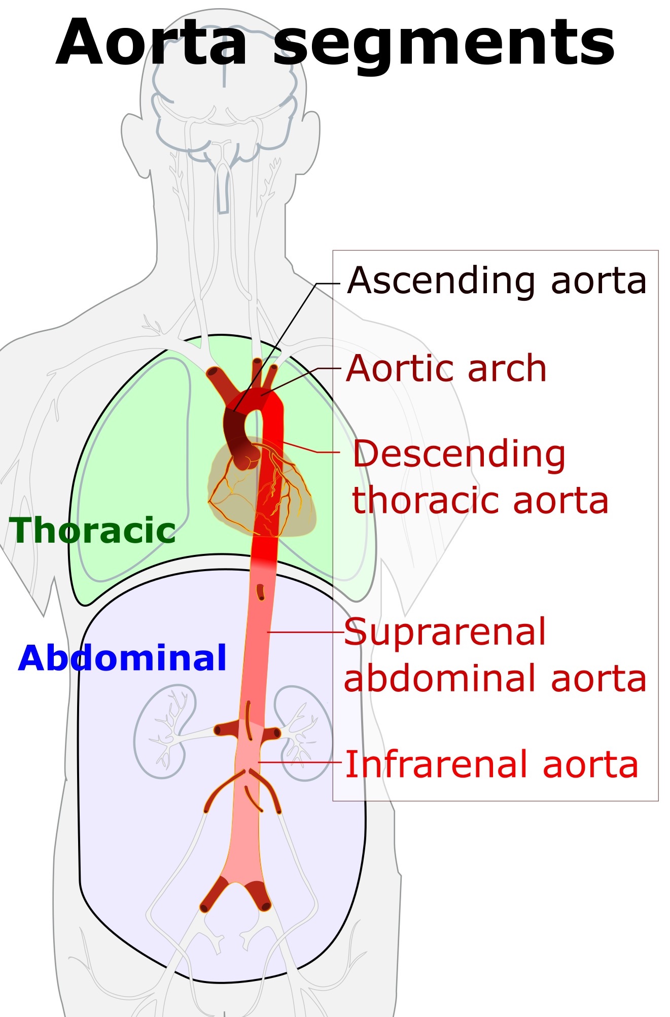
Thoracic aorta branches diagram. As the aortic arch transitions into the thoracic part of the descending aorta it forms a small stricture called the aortic isthmus which is followed by a dilatation. The brachiocephalic trunk the left common carotid artery and the left subclavian artery. A diagram of the aorta. It might be useful to visualize the full aorta as a highway with many potential exits for blood cells vehicles of oxygen.
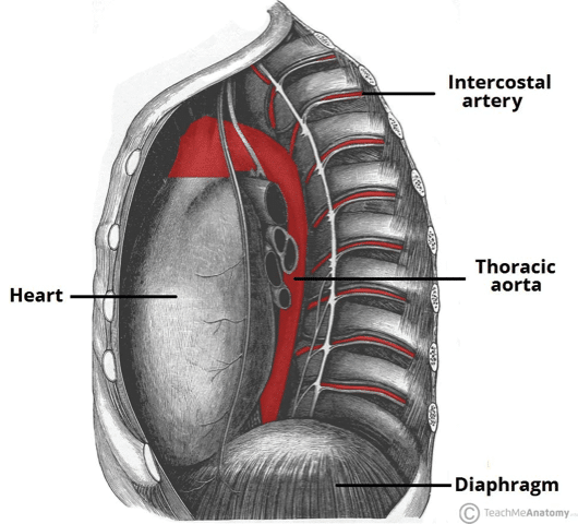
Three arteries branch from the aortic arch and run superiorly figure 1. Proper diagnosis of the problem well in advance helps in early management of the disease. This birth defect causes heart strain due to high blood pressure in the upper body. The descending thoracic aorta is part of the aorta which has different parts named according to their structure or locationthe descending thoracic aorta is a continuation of the descending aorta and becomes the abdominal aorta when it passes through the diaphragmthe initial part of the aorta the ascending aorta rises out of the left ventricle from which it is separated by the aortic valve.
For more anatomy content please follow us and visit our website. Aortic calcification can cause serious illness and its symptoms should not be avoided. The aorta originates from the left ventricle of the heart. If a person complains of any uneasy symptoms medical help should be taken immediately.
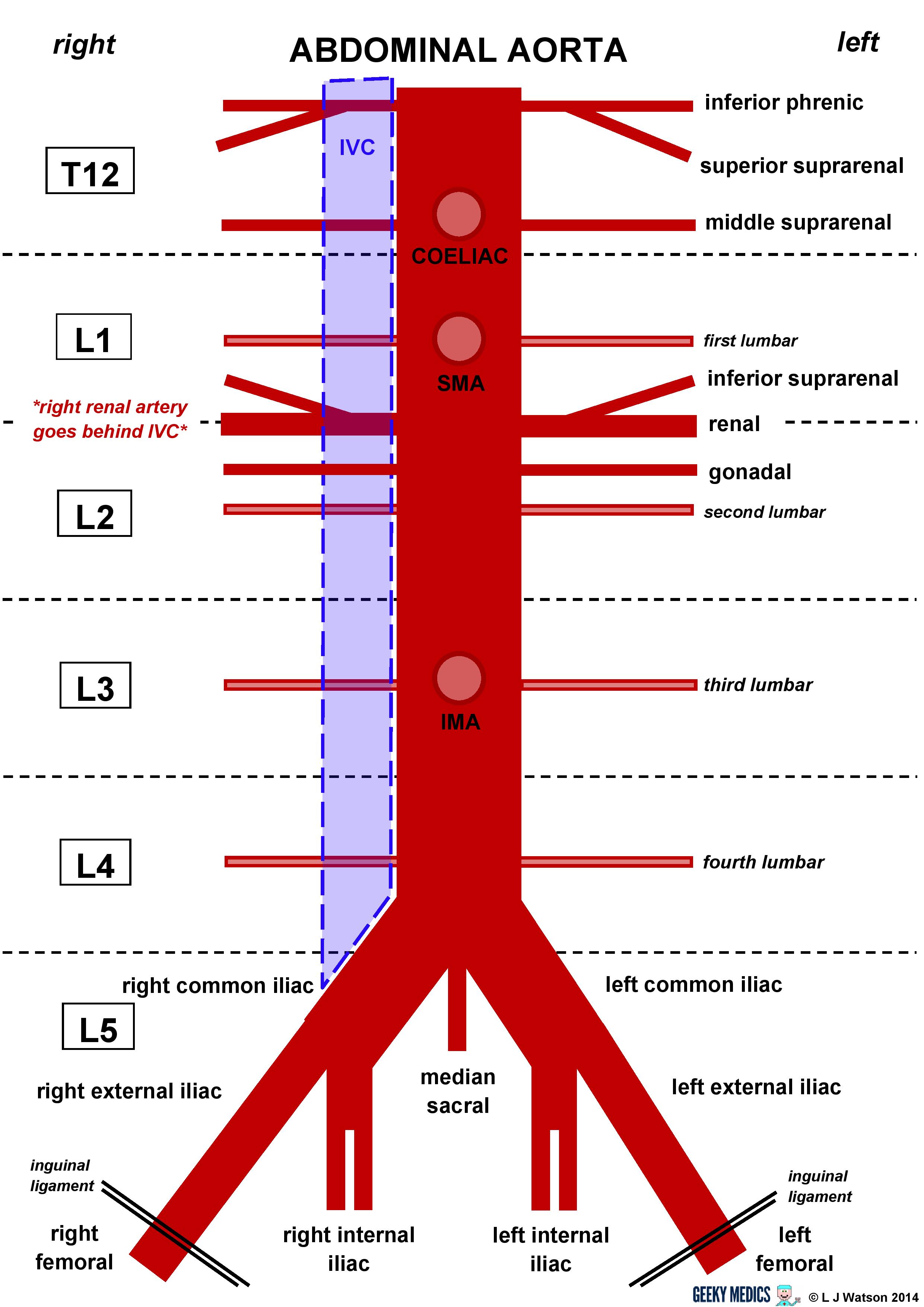
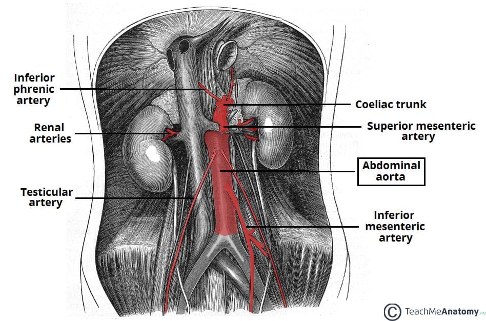
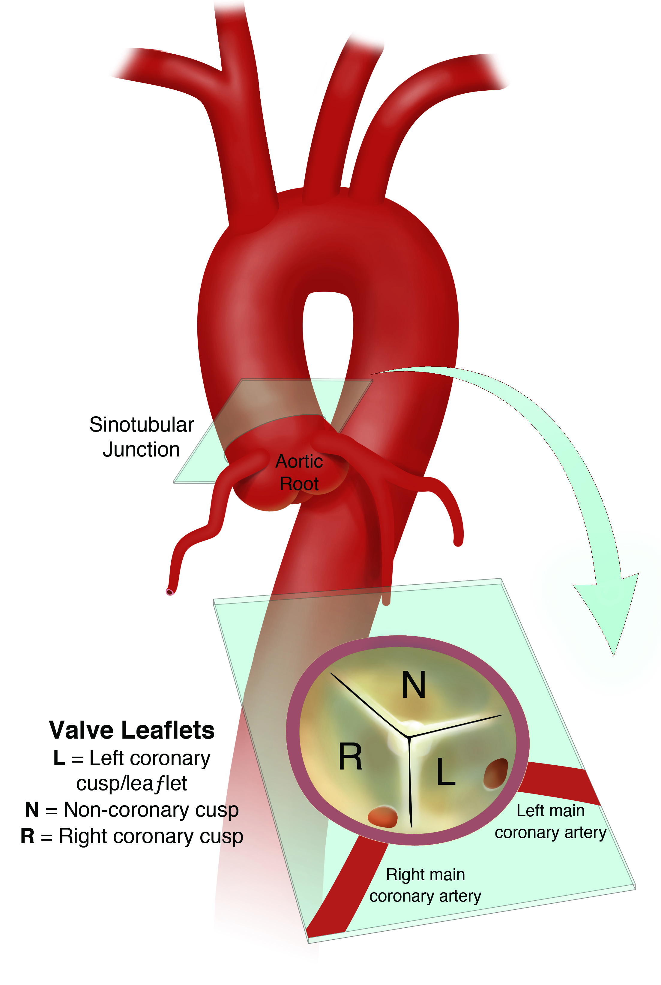
At this level the aorta terminates by bifurcating into the right and left common iliac arteries that supply the lower body. Branches from the. The abdominal aorta is a continuation of the thoracic aorta beginning at the level of the t12 vertebrae. Branches the convexity of the aortic arch gives off three branches.
Narrowing of the aorta between its branches to the arms and those to the legs. From there it travels down the femoral artery in the. The aorta has five separate segments. The brachiocephalic trunk ascends to the right toward the base of the neck where it divides into the right common carotid artery and the right subclavian artery.
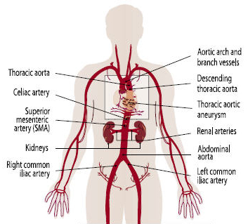
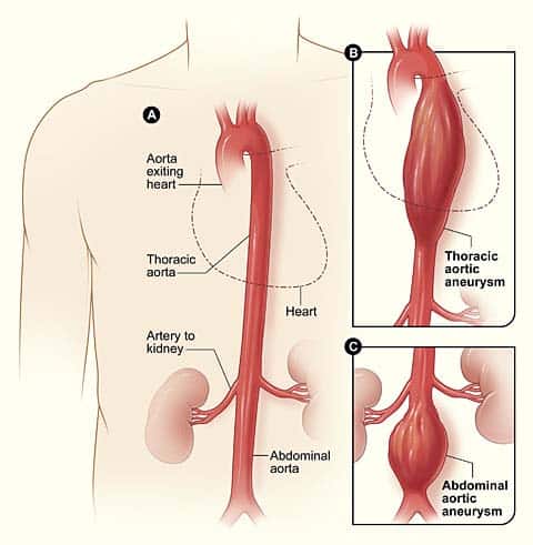
It ends in the abdomen where it branches into the two common iliac arteries. Oxygenated blood leaves the heart and travels down the large thoracic aorta before the aorta divides into two main branches near the abdomen. Calcification of aorta can cause various heart disorders like aortic valve stenosis which blocks the blood circulation to the heart. Coarctation of the aorta.
We are pleased to provide you with the picture named thoracic aorta and abdominal aorta branches diagramwe hope this picture thoracic aorta and abdominal aorta branches diagram can help you study and research.












:background_color(FFFFFF):format(jpeg)/images/library/13386/T5ZCiKoPAprNRZN9BViZqg_Aorta.png)







:background_color(FFFFFF):format(jpeg)/images/library/13387/heart-in-situ_english.jpg)









:background_color(FFFFFF):format(jpeg)/images/library/11095/esophagus-in-situ_english.jpg)


